How the Web Medical Image Viewer can Facilitate Access to Imaging Results
Medical imaging is one of the core elements of medical diagnostics. Most medical centers use it in their everyday work. A frequent solution is to attach a CD or DVD with images obtained during a test to a patient’s results. Images from radiological tests are recorded according to the generally applied medical imaging standard DICOM (Digital Imaging and Communications in Medicine). These images can be displayed in dedicated viewers – DICOM viewers. Multiple solutions used by equipment and software manufacturers cause problems related to the display of the image data saved on carriers, and the need to install many different viewers.

Universal solution assuring quick access to patient image data
Imaging performed in medical centers using equipment from various manufacturers often cannot be opened on many currently available medical image viewers, or, if they can, will be displayed incorrectly. This is, among other things, due to incorrect or varied application of the DICOM standard by equipment and software manufacturers. It also happens that DICOM viewers are saved on the disk with the image data, but operate correctly in just one specified operating system, require installation of additional plug-ins, or are not user-friendly. This makes access to the results troublesome or, in many cases impossible. The functional medical image viewer created by specialists from Comarch Healthcare fully supports the DICOM standard and is compatible with medical images of various origin. The solution was tested by doctors using images of various DICOM types and from many sources. DICOM Runner is a tested viewer that allows test results to be read in many places, providing users with freedom of use.
WWW.DICOMRUNNER.COM – generally available medical image viewer
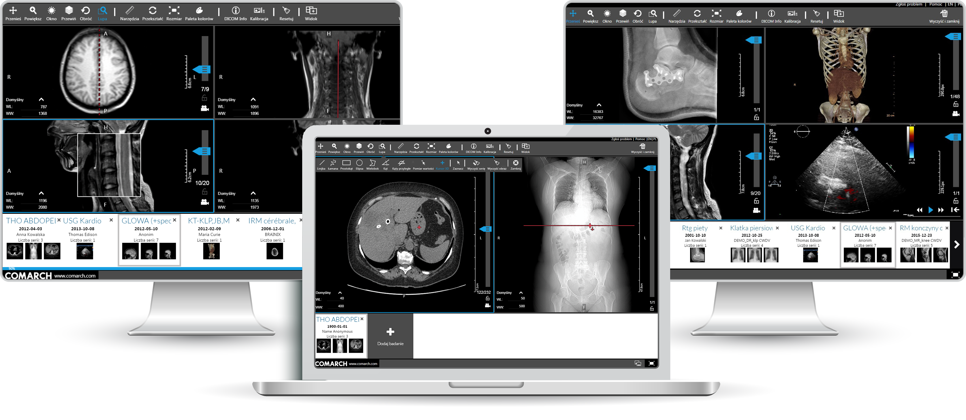
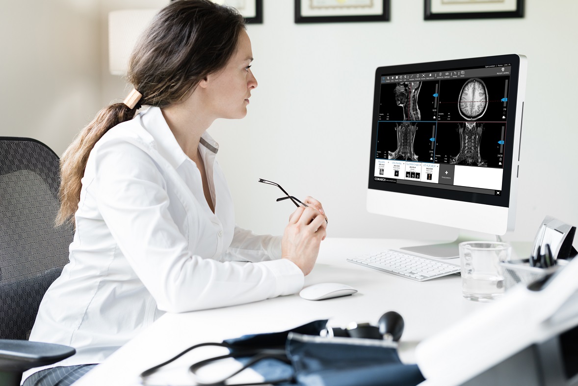
Comarch Healthcare experts have developed a solution to allow radiological test results to be read freely. Comarch DICOM Runner is a tool for the presentation of medical images saved on CD/DVD or other data carriers. The viewer is a class IIa medical product for doctors, physiotherapists, and students of medicine-related sciences, as well as patients and people interested in medical imaging.
The entirely web-based application works quickly, is intuitive, and – most importantly – opens and displays images from various sources correctly. The application allows images generated by various devices, (such as CT, MRI, MG, CR, DR, and PET) to be displayed.
The full version of the application is available online, free of charge, at the address: www.dicomrunner.com

Major Functionalities of Comarch DICOM Runner Include:
- Image processing. The viewer allows any rotation shifting, mirror views, zoom-in and out, and changes to the image size.
- Carrying out various measurements directly on the image. The DICOM Runner user can measure the length of sections and polygonal chains, as well as angles. The viewer also allows regions of interest (ROI) to be marked in the form of rectangles, ellipses, and polygons. Areas are calculated for regions indicated, while for monochromatic images these calculations also include values such as average, standard deviation, and maximum/minimum radiodensity.
- The application allows screen division and simultaneous display of up to four images (1x1, 2x1, 1x2, and 2x2) of various types and from various data carriers, without the need for them to be saved locally. This makes it much easier to compare test results.
- DICOM Runner also allows the location lines that present mutual locations of visible workspaces to be shown. The application provides for the display of two types of location lines – those representing region crossing and those presenting an orthogonal projection of one area compered to another.
- The function of selective synchronization of a study series (e.g. CT or MRI) displayed in particular workspaces allows easy comparison of the images. Synchronized images automatically present the same part of a patient's body, while transformations are propagated onto all synchronized cross-sections. Where there are no location parameters (such as in X-ray imaging), synchronization allows the generation of identical geometric transformations among workspaces.
- The viewer is equipped with a 3D cursor. This tool allows a specific point to be indicated in the patient’s body data space, after which all workspaces on the screen are adjusted in order to present the layer closest to the selected point.
- The application also allows changes to be made to window parameters, such as window width (WW) and window level (WL). The user can change the WW/WL values, and select one of the predefined kits. This option of adjusting the scope of radiodensity mapping onto the greyscale of the displayed image allows visualization of structures of interest to the user. Moreover, if the image has a defined value of interest in Lookup Tables (VOI LUT), it will be displayed by default.
- The viewer features a Multiframe option. This mode is used for reading USG images, allowing sequential images to be played as an animation at a user-selected speed. A series of multi-slice tests can be played automatically in Cine mode.
- By pressing the “DICOM info” button, users can easily view the data in DICOM file headers. Data can be presented in the form of a list with a search option, or as a presentation of major information from the report.
- Considering the security of personal and medical patient data, opening and processing medical images in DICOM Runner occurs locally, on the user side only. Files and image data are not sent via the web, or processed at any place other than on the computer where they are opened.
- As well as offering an intuitive interface, the use of the viewer is made easy owing to a user-friendly manual available at the click of the button in the top right corner.
- In the event of malfunction or error in image data, DICOM Runner displays relevant messages to inform the user immediately about what is happening.
- The viewer allows verification of the conformity of tests displayed with the DICOM standard. Apart from messages about incorrect values in DICOM file headers, an error report is also available.
- The application developed by Comarch Healthcare experts in the web technology is available with interfaces in Polish and English.
An Integral Part of IT Systems for Hospitals
Access to imaging is important not only for radiologists, but also for doctors in other specializations who work on various wards. Comarch DICOM Runner works perfectly well not only as an individual viewer, but also as an integral part of IT systems for hospitals. Owing to cooperation with core medical systems, such as HIS (Comarch Optimed NXT) and systems and imaging labs (Comarch RIS), it provides users with a quick view of medical images. As a part of the Comarch Teleradiology system, it allows the results of imaging from the TeleRIS system level to be viewed across the tele-radiological network.
If you need further information about DICOM Runner, go to dedicated page >>